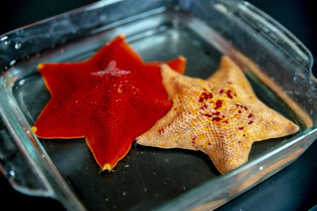
A team of American researchers using the example of starfish studied how living organisms become symmetrical.
All multicellular organisms originate from a single oocyte – the female reproductive cell, the precursor of the egg, which is produced in the ovary during pre-embryonic development. It is she who carries in herself the “plan” for the development of this or that complex organism; it contains a lot of what the organism will become. How this “plan” is created is one of the most important questions in developmental biology.
Scientists from the Massachusetts Institute of Technology (USA) have studied the origin of the so-called initial polarity in the first animal cell, which establishes the axis of symmetry for the developing organism. The work was published in the journal Current Biology. They took the starfish Patiria miniata as an example. These vibrant living organisms exhibit radial symmetry in adulthood: they usually have five axes, but their larvae are bilaterally symmetrical, like humans.
Scientists have come to the conclusion that the mirror symmetry of the larvae is laid while they are in the oocyte stage. And a key role in this process is played by a protein called Disheveled. It is localized at the vegetative or lower end of the oocyte (it is this part of it that defines the posterior end of the embryo) when the cell prepares to divide into two daughter cells.
This protein is a component of a common signaling pathway called Wnt that is found in many creatures in the animal kingdom. It serves a variety of purposes and, in starfish, provides a link between the initial asymmetry of the oocyte and the polarity of the resulting embryo. Wnt is a kind of messenger inside the cells of the starfish: it transmits external signals inward, to the nuclei of the cells.
The scientists used frame-by-frame imaging to see how Disheveled moves around the oocyte as the cell goes through different phases of development. When the starfish oocyte was in a non-dividing phase, the protein was evenly distributed in small clusters throughout the cytoplasm. But as the oocyte was preparing for division, these clusters first dissolved, and then “reappeared” again, but in a different place – at the bottom of the cell, at the farthest point from the nucleus.
This has drawn a clear boundary between the two ends of the oocyte. The same mechanism, according to the researchers, is used not only by sea stars, but, probably, by vertebrates as well. The details of Disheveled’s transformation processes are not yet clear, and scientists intend to study them in the future.
Photo: Starfish Patiria miniata / © phys.org


