3.9 C
Brussels
Tuesday, December 24, 2024
HealthTake a look at the first micrograph of the Omicron strain

Take a look at the first micrograph of the Omicron strain

DISCLAIMER: Information and opinions reproduced in the articles are the ones of those stating them and it is their own responsibility. Publication in The European Times does not automatically means endorsement of the view, but the right to express it.

DISCLAIMER TRANSLATIONS: All articles in this site are published in English. The translated versions are done through an automated process known as neural translations. If in doubt, always refer to the original article. Thank you for understanding.

Newsdesk
Newsdeskhttps://europeantimes.news
The European Times News aims to cover news that matter to increase the awareness of citizens all around geographical Europe.

Scientists from the University of Hong Kong managed to take a photo of the new omicron coronavirus strain.

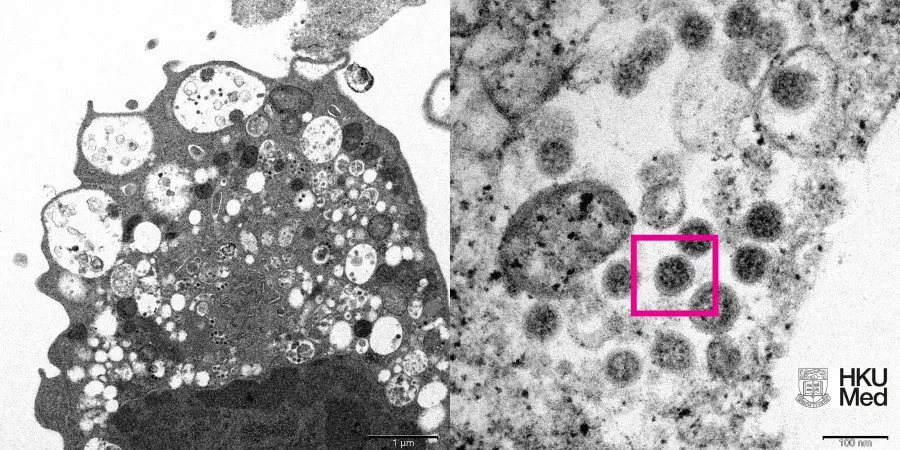
The photograph shows an electron micrograph of a monkey kidney cell (Vero E6) infected with an omicron. There is a picture at low and high magnification.

The photo was published on the website of the medical faculty of the university.

The picture shows lesions with swollen vesicles, which contain viral particles. At high magnification, the researchers were able to make out clusters of characteristic spherical objects with crown-shaped spikes on their surface.

The day before, on December 6, it became known that the first two cases of infection with the omicron strain were confirmed in Russia. These are two people who came from South Africa.

Earlier, “Hi-Tech” wrote about what is already known about the omicron-strain and how dangerous it is.

- Advertisement -

More from the author

- EXCLUSIVE CONTENT -spot_img
- Advertisement -
- Advertisement -
- Advertisement -spot_img
- Advertisement -

Must read

Latest articles

- Advertisement -